
Cell Structure & Transport: Why This Chapter Feels Like the Heartbeat of AP Biology
Welcome—lean in. If you’re studying cell structure and transport for AP Biology, you’re not alone: this section connects nearly every other unit in the course. From understanding how organelles coordinate life-sustaining chemistry to predicting how a cell responds to its environment, mastery here gives you mileage across the entire exam.
This post offers evidence-based explanations (not just memorization tricks), clear analogies, exam-style examples, and practical study moves you can use immediately. Along the way I’ll highlight study strategies, sample calculations, and why a tailored approach—like Sparkl’s personalized tutoring with 1-on-1 guidance and tailored study plans—can accelerate your understanding.
How to Read This Guide
Think of this as a toolkit. Each section pairs an accessible explanation with the evidence you’ll need to justify answers on free-response questions (FRQs) and multiple choice. After reading, you should be able to: (1) describe structures and functions, (2) predict transport outcomes, and (3) justify those predictions using scientific reasoning.
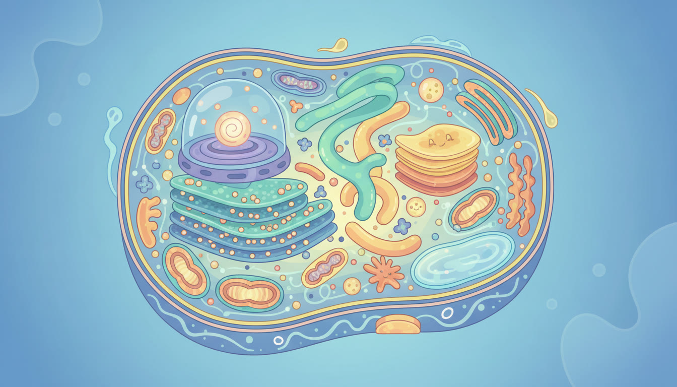
1. Cell Architecture: Structure Followed by Function
Cells are complex but organized. AP exam questions often test not only identification of organelles but the reasoning behind why particular structures enable specific functions. Below is a concise map of the major players and the evidence that connects form to role.
Key Organelles and Evidence-Based Roles
- Nucleus: Stores and protects DNA; nuclear pores regulate transport of RNA and proteins. Evidence: nuclear pore size and active transport mechanisms explain selective movement of large molecules.
- Ribosomes: Sites of protein synthesis. Evidence: polyribosome arrays and mRNA association correlate with high translation rates.
- Rough Endoplasmic Reticulum (RER): Co-translational insertion and folding of secreted/membrane proteins. Evidence: ribosomes attached to RER and signal peptide-dependent targeting.
- Smooth ER: Lipid synthesis and detoxification. Evidence: cells active in steroid synthesis show abundant smooth ER.
- Golgi Apparatus: Modifies, sorts, and ships proteins. Evidence: vesicle trafficking and glycosylation patterns change protein destination and function.
- Mitochondria: ATP production via oxidative phosphorylation. Evidence: presence of electron transport chain complexes in inner membrane and correlation between cell energy demand and mitochondrial abundance.
- Lysosomes: Digestive compartments for macromolecules. Evidence: low internal pH and hydrolytic enzymes enable degradation of cellular debris and ingested particles.
- Peroxisomes: Oxidative reactions that break down fatty acids and detoxify. Evidence: catalase presence and H2O2 metabolism confirm function.
- Plasma Membrane: Selective barrier; houses receptors and transporters. Evidence: differential permeability to ions/molecules and presence of membrane proteins mediating signal transduction.
Structural Themes That Repeat
- Membrane compartments—compartmentalization increases efficiency and prevents cross-reaction of pathways.
- Surface area to volume tradeoffs—many organelles have folded membranes (cristae, thylakoids, RER) to maximize reaction space.
- Localization of processes—steps of a pathway are often physically co-located (e.g., glycolysis cytosolic, oxidative phosphorylation mitochondrial).
2. Membrane Structure: The Bilayer as a Dynamic Marketplace
The fluid mosaic model remains the best explanatory framework. It emphasizes a lipid bilayer matrix with embedded proteins that move laterally, creating a dynamic surface for transport, signaling, and cell recognition.
Core Components and Why They Matter
- Phospholipids: Amphipathic; their hydrophobic tails and hydrophilic heads drive bilayer formation and create a semipermeable barrier.
- Cholesterol: Modulates membrane fluidity; higher cholesterol reduces fluidity at high temperatures and prevents solidification at low temperatures.
- Integral proteins: Channels, carriers, receptors—mediate selective transport and signaling.
- Peripheral proteins and glycocalyx: Often involved in cell recognition and membrane support.
Evidence Students Should Be Able to Use
Experimental support comes from freeze-fracture microscopy (which shows protein distribution), fluorescent recovery after photobleaching (FRAP) for lateral mobility, and permeability assays demonstrating differential passage of polar vs nonpolar molecules. When you explain a transport phenomenon on the AP exam, cite the physical property: size, charge, and polarity determine passive permeability.
3. Passive Transport: Diffusion, Osmosis, and Facilitated Diffusion
Passive transport moves substances down their electrochemical gradients without cellular energy input. The AP exam likes to test whether you can predict net movement and explain the underlying driving forces.
Diffusion and Concentration Gradients
Diffusion is driven by kinetic energy and the tendency toward equilibrium. Small nonpolar molecules (O2, CO2) and lipophilic molecules cross the membrane easily. For polar or charged molecules, the bilayer is a barrier.
Osmosis: A Special Case of Water Diffusion
Osmosis is the net movement of water across a selectively permeable membrane toward the compartment with higher solute concentration (lower water potential). On the AP exam, expect questions that ask you to determine whether a cell will swell, shrink, or stay the same in a given solution.
| Solution Type | Relative Solute | Cell Response | Example |
|---|---|---|---|
| Hypotonic | Lower external solute | Cell swells (possible lysis in animal cells) | Freshwater around animal cell |
| Isotonic | Equal | No net change | Most cells in vertebrate blood plasma |
| Hypertonic | Higher external solute | Cell shrinks (plasmolysis in plant cells) | Saltwater around freshwater cell |
Facilitated Diffusion
Carrier and channel proteins allow polar or charged substances to cross down their gradient. Evidence includes saturation kinetics for carrier proteins and gated channels responding to stimuli (voltage, ligand, mechanical force).
4. Active Transport and Electrochemical Gradients
Active transport moves substances against their gradient and requires energy—often ATP. The sodium-potassium pump (Na+/K+ ATPase) is the classic AP example: it maintains membrane potential and ionic balance by exporting Na+ and importing K+.
Why Gradients Matter
- Gradients store potential energy (electrochemical potential) used for secondary active transport (cotransport).
- Many cellular processes—nerve signaling, nutrient uptake, and pH regulation—depend on pumps and transporters.
Example: Secondary Active Transport
Cells often use the energy of an existing Na+ gradient established by Na+/K+ ATPase to drive uptake of glucose against its concentration gradient via a cotransporter (symporter). This indirect use of ATP is an elegant economy of energy that appears frequently in FRQs.
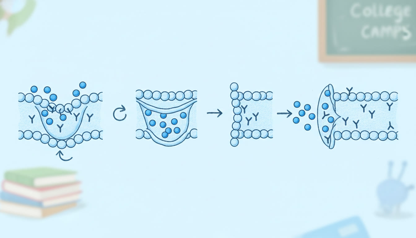
5. Bulk Transport: Endocytosis and Exocytosis
Large molecules, particles, or large volumes of fluid use membrane remodeling mechanisms:
- Endocytosis: Phagocytosis (cell eating), pinocytosis (cell drinking), and receptor-mediated endocytosis (specific uptake via clathrin-coated pits).
- Exocytosis: Vesicle fusion to release contents or to insert proteins/lipids into the plasma membrane.
Evidence to reference: presence of coated pits, vesicle tracking in live-cell imaging, and the role of SNARE proteins in vesicle fusion.
6. Putting It Together: Example FRQ Walkthrough
Let’s practice with a short hypothetical FRQ-style scenario (common on AP tests). Read it and use the evidence-based reasoning below to craft an answer in your head:
Scenario: A liver cell is exposed to a toxin that disrupts the function of the Golgi apparatus. Predict three cellular consequences and explain the mechanistic evidence for each prediction.
Model Answer (Evidence-Backed)
- Protein misprocessing and secretion defects: The Golgi modifies and sorts proteins. Without it, secreted proteins may be improperly glycosylated or fail to be packaged into secretory vesicles, reducing secretion. Evidence: Changes in glycosylation patterns alter protein stability and targeting.
- Accumulation of vesicles or proteins in the RER: Proteins synthesized into the RER will not progress through secretory pathway efficiently, causing ER stress and potentially triggering the unfolded protein response. Evidence: Cells with blocked Golgi show increased ER chaperone expression and dilated ER cisternae under electron microscopy.
- Disruption of membrane protein trafficking: Membrane proteins needing Golgi-mediated modifications will mislocalize, altering membrane transport and signaling. Evidence: Mislocalization can be observed by immunofluorescence and correlates with functional deficits (e.g., reduced receptor-mediated endocytosis).
Note how each prediction pairs structure, expected functional outcome, and a testable piece of evidence—this is the kind of logic AP graders expect.
7. Study Strategies That Actually Work (Evidence-Based)
High-yield content matters, but approach matters more. Use these strategies to retain concepts and deploy them under test pressure.
Active Recall and Spaced Practice
- Make concise question cards that ask for mechanism and evidence (e.g., “How does Na+/K+ ATPase contribute to secondary active transport? Give an experimental observation that supports this.”).
- Practice these cards in spaced intervals—evidence supports better retention than massed review.
Explain to a Peer or Tutor
Teaching a concept forces you to supply causation and evidence. If you can explain membrane transport so a friend can sketch gradients and explain net flux, you’re ready. Personalized tutoring—such as Sparkl’s 1-on-1 guidance—can give targeted feedback when you try to teach back, honing misconceptions and accelerating mastery.
Use Diagrams and Quantitative Reasoning
Draw gradients as vectors and practice small calculations (osmolarity, relative concentrations). Many students miss points by failing to quantitatively justify direction of net flux—learn to show your work concisely.
Practice FRQs and Retrospective Revision
After each practice FRQ, write a brief note on what evidence you used and which pieces of the prompt demanded that evidence. Over time you’ll build an internal rubric for answer completeness.
8. Common Pitfalls and How to Avoid Them
- Confusing diffusion with active transport: If a process requires ATP or moves against a gradient, it’s active. Always ask: is energy required?
- Ignoring membrane permeability rules: Don’t assume all small molecules cross freely—polarity and charge matter.
- Overgeneralizing plant vs animal responses: Plant cells have a cell wall, changing the outcome in hypotonic solutions (turgor pressure vs lysis in animal cells).
- Missing evidence in FRQs: State the experimental or structural basis behind your prediction—link mechanism to a testable sign.
9. Quick Reference Table: Transport Mechanisms at a Glance
| Mechanism | Energy Required? | Example Proteins | AP-Style Evidence |
|---|---|---|---|
| Simple Diffusion | No | None | Permeability assays showing faster equilibration for nonpolar gases |
| Facilitated Diffusion | No | Ion channels, GLUT transporters | Saturation kinetics, channel gating studies |
| Primary Active Transport | Yes (ATP) | Na+/K+ ATPase, Ca2+ pumps | ATP hydrolysis assays and ion gradient measurements |
| Secondary Active Transport | Indirect (uses existing gradients) | Symporters, Antiporters | Disrupted gradient abolishes coupled transport (experimental evidence) |
| Endocytosis/Exocytosis | Yes (membrane remodeling; often ATP) | Clathrin, SNARE proteins | Microscopy tracking of vesicle dynamics |
10. Bringing Evidence Into Your Answers
AP graders reward answers that connect biological structure to measurable outcomes. When you make a claim, back it with a reason that could be experimentally observed. For example:
- Claim: “Na+/K+ ATPase helps maintain membrane potential.”
- Evidence: “Blocking the pump with ouabain reduces the membrane potential magnitude as measured by intracellular electrode recordings.”
- Explanation: “Because fewer positive charges are exported, the electrochemical difference across the membrane collapses, impacting secondary transport.”
That three-part structure (claim, evidence, explanation) improves clarity and the scientific rigor of your response.
11. How Personalized Tutoring Can Accelerate Mastery
General textbooks and videos are useful, but targeted practice on the exact mistakes you make produces faster gains. Tutoring that combines subject expertise with personalized plans—like Sparkl’s personalized tutoring offering expert tutors and AI-driven insights—can help you identify conceptual gaps, prioritize high-yield topics, and get focused feedback on FRQs. Use it to rehearse explanations aloud, practice evidence-based answers, and refine timing on the exam.
12. Final Checklist Before the Exam
- Can you sketch and label a typical animal and plant cell and explain organelle functions with evidence?
- Can you predict the direction of net water and solute movement in hypothetical scenarios and justify your answer experimentally?
- Are you comfortable converting a descriptive prompt (e.g., “membrane becomes more permeable to Na+”) into mechanistic predictions and experimental observations?
- Have you practiced FRQs using the Claim-Evidence-Reasoning structure?
Closing: Turn Curiosity into Exam Confidence
Cell structure and transport is where molecular logic meets experimental evidence. When you approach each concept asking “What would I observe if this changed?” you start answering at the level AP graders value most. Use diagrams, practice problems, evidence-based reasoning, and targeted help where needed. If you want a customized plan—a few practice prompts tuned to your weak spots and 1-on-1 feedback—consider a short session of personalized tutoring to accelerate progress. Sparkl’s tailored study plans and expert tutors can be a smart boost if you’re short on time.
Study actively, test with the lens of evidence, and keep your explanations crisp. You’ve got this—cells run on logic, and so can your exam answers.


No Comments
Leave a comment Cancel